


It was late 2014 when Phil Torres first showed me the photos from his recent trip to the Peruvian Amazon. Among them were amazing images of the tropical wildlife, from brilliant macaws to elusive pumas. But there were a few critters in that album that stood out to us in particular. Flipping through his camera, Phil said something to the effect of, "Check out this butterfly dude, it hangs out with ants on bamboo."
You can watch the video Phil and I put together on this discovery
After Phil showed me a few more photos, it was clear that this butterfly-ant interaction was not due to chance...something was going on here.
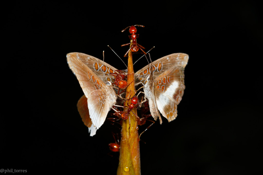
Multiple butterflies (both male and female) gathering on a bamboo stalk in the presence of ants.

Up-close shot of the butterflies and ants feeding from the sap secretions emitting from the bamboo shoot.
Phil and I both have backgrounds in entomology, and yet we had never seen anything like this before. I mean sure, we knew that some butterfly larvae have symbiotic relationships with ants, known as myrmecophily. This is well documented, as many of the caterpillars that associate with ants have special organs that secrete sugars and amino acids. The ants get a sugary nutritious meal from the caterpillars and, in return, the fragile caterpillars get personal ant bodyguards which defend against predators and parasites. But this is not the case for the adult butterflies, which usually have to evade ants, lest they become their next meal.
One more thing, Phil said, "Look at the three red spots on the butterfly wing. Kind of looks like the ants they're with on the bamboo. Maybe it's some sort of mimicry."
Alright, now I was really interested. The butterfly appears to be a known species, Adelotypa annulifera, but these pictures could be revealing an undocumented observation for this butterfly interacting with ants and a potentially new wing-mimicry pattern. Super cool, I thought, but there was just one problem: we know little about this butterfly beyond some dead pinned specimens. What is its life cycle? Where do the larvae develop? What do the larvae even look like? In other words, next to nothing was known about the life history of this butterfly. So to solve this mystery, Phil and I decided to collaborate. I was making a return trip to this exact field site in the coming months, so I set out to uncover the missing pieces of this puzzle.
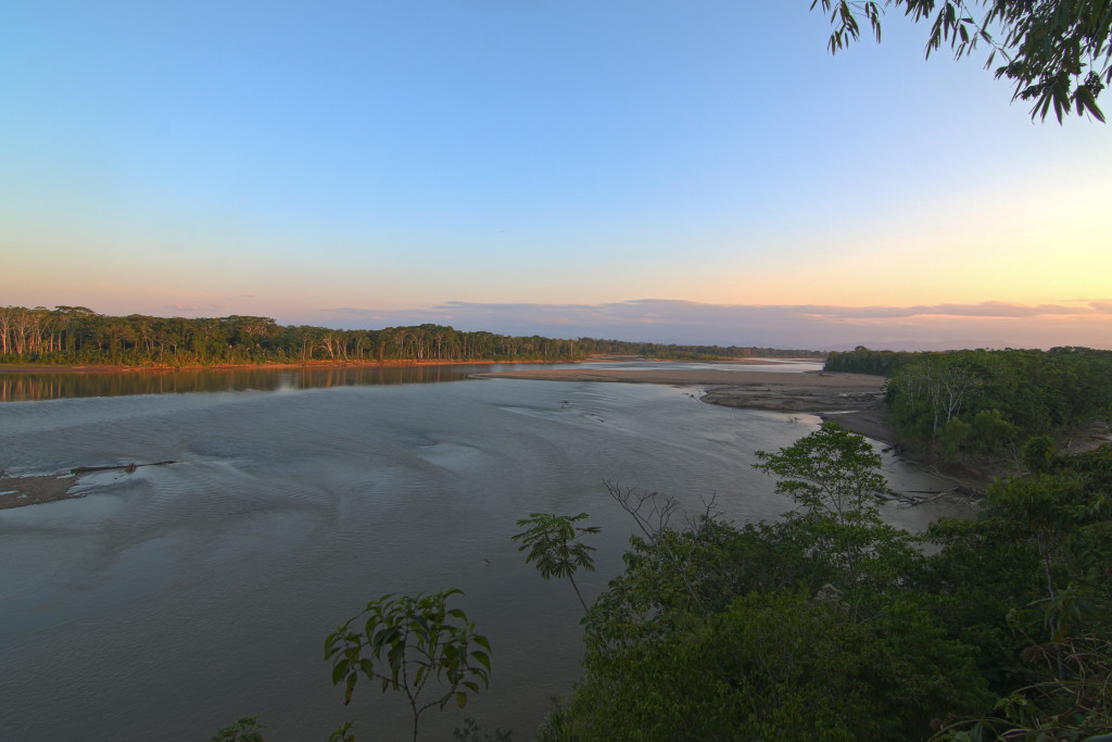
Overlook of the Tambopata River at our field site in Southeastern Peru.
The challenge with this type of fieldwork is that the Amazon rainforest is very big, and the critters we are looking for are very small. Since Phil observed the butterflies on bamboo, I ventured out to the same habitat, a trek from the Tambopata Research Center. I recall the jungle seemed particularly hot, humid, rainy, and muddy during that expedition, but I was determined to find our caterpillars and butterflies.
After hours of hiking through the Peruvian Amazon and getting continuously soaked by rain downpours, I found myself in the bamboo forest where we knew our butterflies liked to hang out. After checking dozens of bamboo plants, things seemed futile, as I wasn't finding any signs of our butterflies of interest. But persistence is the key to field work. I then saw a young bamboo shoot poking out of the mud, and noticed a leaf near the base of the bamboo, close to the ground.

Bamboo stalk with a leaf wrapped around the shoot near the base of the plant.
I pulled the leaf back and to my utter shock, found myself staring directly at two caterpillars nestled against the bamboo, and an agitated ant hovering over the Lepidoptera larvae. My heart was pounding - did I really just find our caterpillars in this vast rainforest!? Clearly they were myrmecophilous, as the ant was trying to protect them.
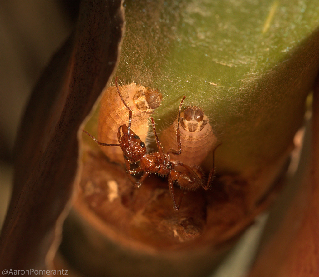
First observation of the caterpillars with an ant bodyguard.
Although excited from the find, I knew the job wasn't done. This could be any species of caterpillar, so I knew I had to watch them turn into pupae and finally into adults in order to truly confirm that these belonged to the same butterfly species. I continuously checked up on the caterpillars at this spot and took numerous photos and video. After a couple of days, I found our little critters in the same location, but this time they had transformed into pupae! At this point, I gently collected them and brought them to a small insect cage at the Tambopata Research Center to see if they would emerge as butterflies. I had my fingers crossed, hopefully they would survive to adulthood.
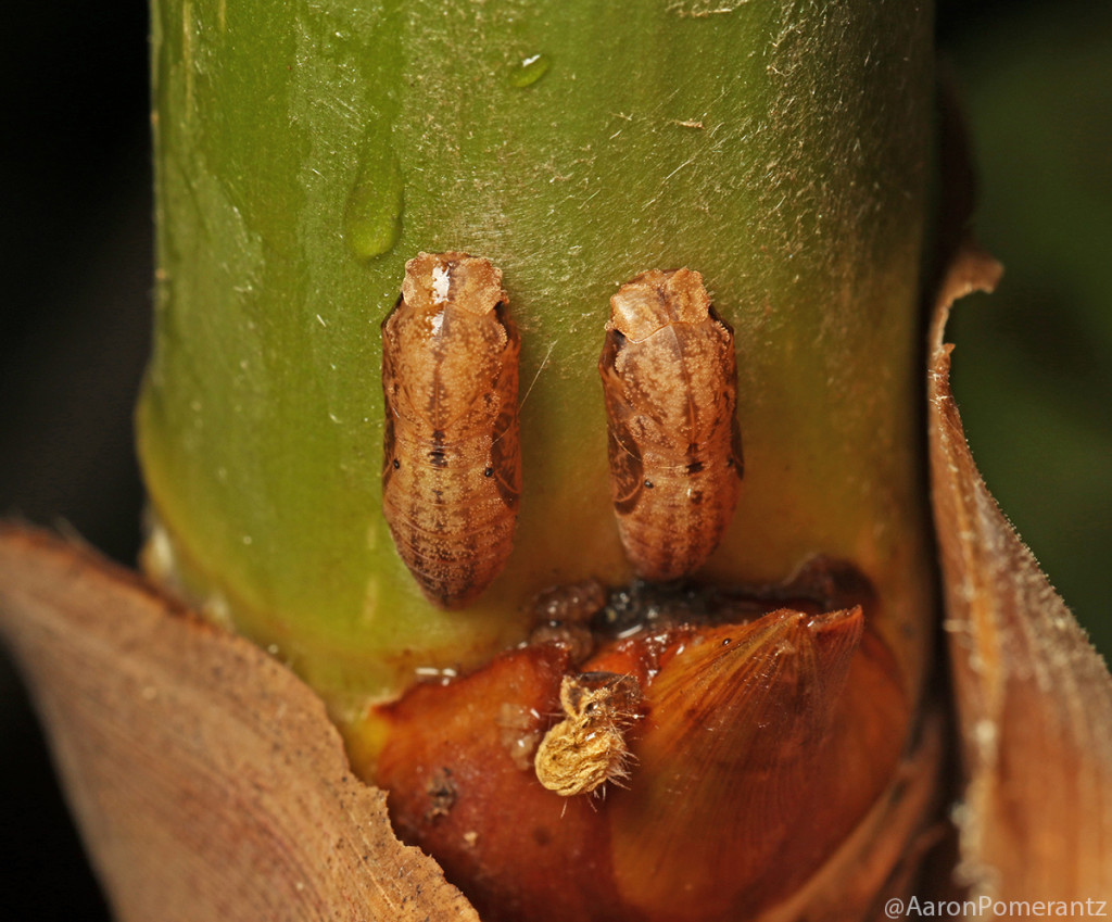
The caterpillars later turned into pupae.
Fast forward, and one day I walked past the little insect cage when I noticed some activity. Wings fluttering. One of the pupae had successfully eclosed! So the moment of truth, what butterfly was it? My jaw dropped when I noticed it was, in fact, the same butterfly (Adelotypa annulifera) that Phil had taken pictures of previously. What this means is, we had just completed the entire life cycle of the butterfly, from egg to larvae to pupae, and finally adult. We now felt that we had enough material to write this up as an official scientific publication.

Figure 1 from the publication, the immature life stages of the butterfly and their association with ants. (A) Eggs with Megalomyrmex ant, (B) First instar larva with Ectatomma tuberculatum ant (C) Mid-instar larva with Pheidole ant (D) Mid-instar larvae with bullet ants (E) Final instar larva with E. tuberculatum ant (E) Pupae.
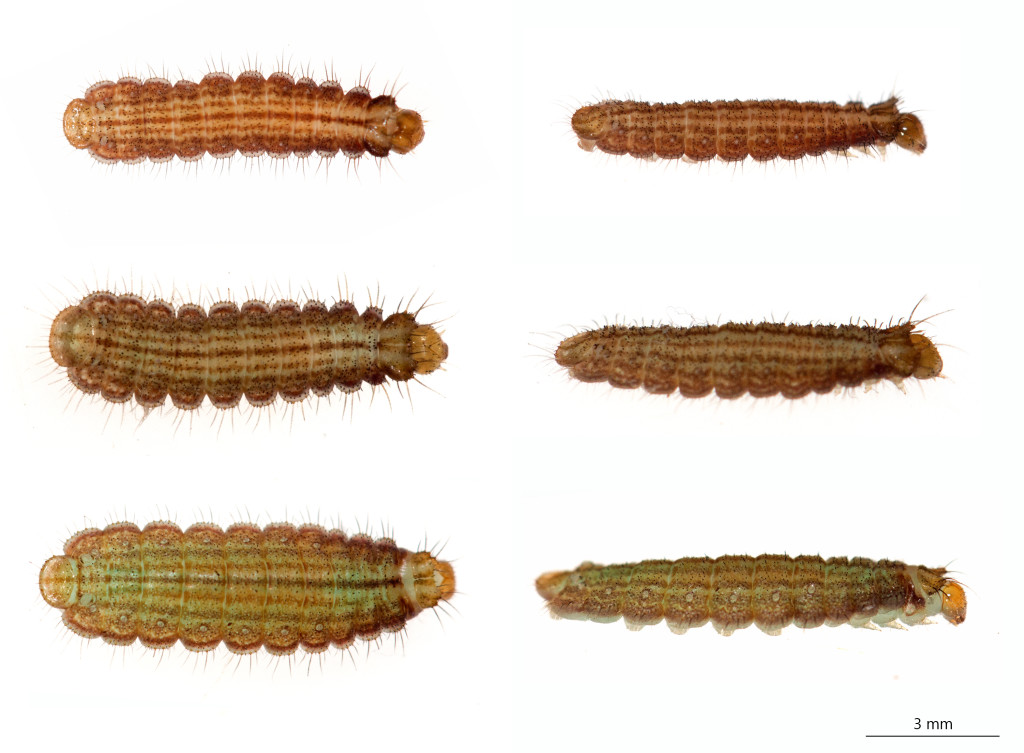
Figure 2. Dorsal (left) and lateral (right) views of early instar caterpillar.

Figure 3. Final instar larva (left) and pupa (right).

Figure 4. Adult Adelotypa annulifera interactions with ants on bamboo. (A) Ants touching the butterfly wings with antennae. (B) Ant crawling on butterfly wing. (C) Ants touching butterfly abdomen. (D-E) Butterflies and ants utilizing extrafloral nectary resources on bamboo. (F) Butterfly drinking bamboo fluid from the ant.

Figure 5. The butterflies and their putative wing pattern mimicry. (A) male butterfly (left) and female butterfly (right) perched on bamboo shoot in presence of red ants. Views of the wing pattern (B) ventral (C) dorsal and (D) lateral.
Overall, it was really exciting collaborating with Phil to discover and publish this life history, which is completely new for this genus of butterflies. In addition, we think the fact that butterflies steal a resource from the ants and let the ants crawl all over them indicates that some complex chemical signaling is going on. Perhaps the butterflies are utilizing a pheromone from their larval stage, potentially allowing the butterfly to take advantage of the ants, which would normally tear a fragile butterfly to shreds. The three red spots on the butterfly wing also look strikingly like the red ants (at least to us) and perhaps serve as a form of mimicry (if a butterfly looks like red ants that bite and sting, a bird may be less inclined to eat it). However, it should be noted that these are just our hypotheses at the moment and, like any hypothesis, should be rigorously tested before we can claim to back it up. We hope to do so, because there most certainly seems to be more to this incredible tropical butterfly than meets the eyes. Stay tuned...

Phil and I enjoying a boat ride along the river in the Amazon rainforest.
For more info, you can download the PDF here: Torres_Pomerantz_Adelotypa_Publication_2016
One more fun fact: it was actually during this trip that I accidentally discovered a totally new, yet unrelated, butterfly-ant relationship. The jungle is full of endless surprises -
[embed]https://www.youtube.com/watch?v=Z1a4zIjo3uU[/embed]
Mystery of the Yellow Bulbs, video and blog post http://blog.perunature.com/2015/11/mystery-of-yellow-bulbs-discovery-of.html
-Aaron
My Instagram: @NextGenScientist & Twitter: @AaronPomerantz
Phil's Instagram: @phil_torres & Twitter: @phil_torres


